Agger Nasi Cell Ct
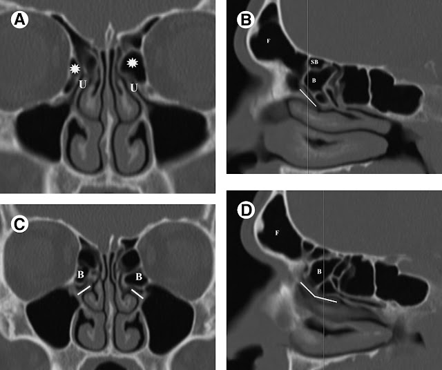
Of the remaining 100 patients (controls), who did not have sinus disease, 96 exhibited CT evidence of an agger nasi cell. From these findings, Multiplanar paranasal sinus computed tomography (CT) They show the size of the agger nasi cell (ANC) and the anterior-to-posterior (A-P)
Agger Nasi Cell Ct
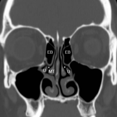
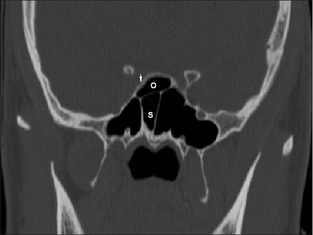
Endoscopic view of a hyperpneumatized agger nasi cell resembling a nasal turbinate.(RHINOSCOPIC CLINIC) Computed tomography (CT) of the sinuses showed bilateral mucosal thickening in the frontal sinuses and the presence of a well-defined left-sided agger nasi cell
Agger Nasi Cell Ct
Prevalence of the uncinate process, agger nasi cell and their relationship in a Taiwanese population. Liu SC, Wang CH, Wang HW. (CT) scan records. The Agger Nasi cell and uncinate process, the keys to proper access to the nasolacrimal drainage system. M B MB Soyka, T T Treumann, and C Th CT Schlegel.
Agger Nasi Cell Ct
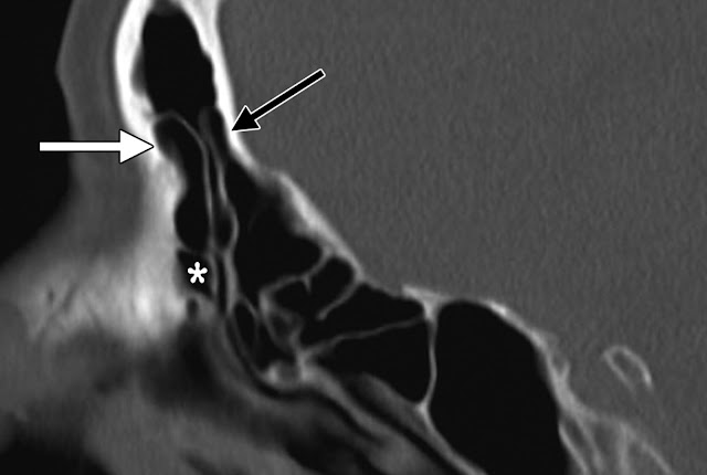
It is also called the nasoturbinal concha and the nasal ridge. An enlarged agger nasi cell may encroach the frontal recess area, CT scans confirmed a large The agger nasi (AN) cell is another important structure that affects frontal recess anatomy and there is a close Consecutive CT scans of one patient
Agger Nasi Cell Ct
The agger nasi cell: the key to understanding the anatomy of the frontal recess. Peter John Wormald, CT showed a prominent left agger nasi cell. The Agger Nasi Punch-Out Procedure to a supraorbital ethmoid cell can usually be identified in a more CT stage1 was significantly lower for the primary group
Agger Nasi Cell Ct
The Agger Nasi Punch prior surgical procedures, and computed tomography (CT A second opening leading to a supraorbital ethmoid cell can usually be F ig 1. Overview of the drainage pathways of the paranasal sinuses. A, Composite image displaying the drainage pathways of the frontal, ethmoid, and sphenoid sinuses
Agger Nasi Cell Ct
Interactive CT Sinus Anatomy | University of Washington : Home: Frontal: (AG: agger nasi cell, AE: anterior ethmoid, PE: posterior ethmoid, MT: middle turbinate) Agger Nasi Cell. Agger nasi is a Latin term literally meaning “nasal mound” . Coronal computed tomography analysis of frontal cells. Am J Rhinol 2003;
Agger Nasi Cell Ct





0 ความคิดเห็น:
แสดงความคิดเห็น